Placental Abnormalities
The placenta is an organ that your body grows during pregnancy. For an overview of its structure, development, and function, please see our handout “The Placenta“. The placenta is highly vascular, meaning there is lots of blood flow. Abnormalities in the placental location and/or how the placenta is attached to the birthing parent’s body can put the patient at risk for excessive bleeding which can be dangerous for both the patient and fetus.
This handout covers some abnormalities that can affect the placenta. These conditions are relatively uncommon, and can include placenta previa, vasa previa, and placenta accreta spectrum disorders.
Placenta Previa
Placenta previa and low-lying placenta are disorders of placental location in relation to the cervix. Placenta previa describes when the placenta is completely covering the opening of the cervix. Low-lying placenta refers to when the placenta is within 2cm of the cervix. The cervix is a tubular structure that connects the uterus to the vagina. It is dangerous for the placenta to be close to the cervix because small, normal changes to the cervix throughout pregnancy or near labour can cause excessive bleeding which can be dangerous for the birthing parent and fetus.
Diagnosis & Symptoms
Placenta previa and low-lying placenta is diagnosed by transvaginal ultrasound conducted at 18-21 weeks. The majority of placenta previa and low-lying placenta resolves as the uterus continues to grow in pregnany (the placenta moves away from the cervix). Therefore, the diagnosis is reconfirmed by ultrasound in the third trimester. Placenta previa is either asymptomatic (no symptoms) or presents with painless vaginal bleeding.
Implications
Placenta previa puts you at risk of antepartum hemorrhage (bleeding), hospitalization, blood transfusion, postpartum hemorrhage, and preterm birth. You are also at higher risk of placenta accreta spectrum, especially if your placenta is anterior (on the front wall of the uterus, facing your belly). You should present to the hospital if you experience any vaginal bleeding.
People with placenta previa can continue their daily activities and engage in light exercise, but they should avoid vaginal and anal sexual activity, or inserting objects into the vagina or anus.
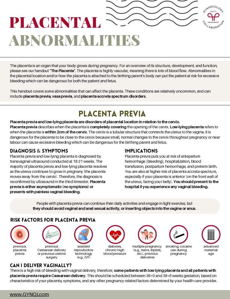
Risk Factors for Placenta Previa
- Previous placenta previa
- Previous Caesarean delivery or previous uterine surgery
- Assisted reproductive (e.g., IVF)
- Diabetes, chronic high blood pressure
- Multiple pregnancy (e.g., twins, triplets, etc.)
- Smoking, cocaine use during pregnancy
- Advanced maternal age
Can I Deliver Vaginally?
There is a high risk of bleeding with vaginal delivery, therefore, some patients with low lying placenta and all patients with placenta previa require Caesarean delivery. This should be scheduled between 36+0 and 38+6 weeks gestation, based on characteristics of your placenta, symptoms, and any other pregnancy-related factors determined by your health care provider.
Placenta Accreta Spectrum (PAS)
Placenta accreta spectrum (PAS) is a term used to describe a spectrum of disorders related to when the placenta (the organ that provides nutrients/oxygen and removes waste from the developing fetus) attaches too deeply into the uterus wall. PAS disorders make it difficult to for the placenta to detach from the uterus after delivery and can lead to significant bleeding.
PAS is rare, affecting 1 in 500 pregnancies. The incidence is rising, likely because of an increasing incidence of Caesarean delivery, which increases the risk of PAS.
PAS is typically asymptomatic, and is diagnosed with imaging techniques including ultrasound and MRI. It often co-exists with placenta previa and may present with vaginal bleeding.
In the most severe cases, this can be very dangerous for the birthing parent and the baby. Possible risks include the need for a blood transfusion, requiring preterm delivery less than 37 weeks, prolonged hospitalization (before and after delivery), emergency hysterectomy (removing your uterus), increased surgical complexity (meaning higher risk of surgical injury to surrounding organs), higher risk of blood clot in the legs or lungs, higher risk of postpartum infection, or very rarely, death. Although this can be scary, patients with PAS who are diagnosed early and have a planned delivery have very good outcomes.
Various Types of PAS
In order of severity, the PAS disorders includes placenta accreta, placenta increta, and placenta percreta. They differ in how deeply the placenta implants into the wall of the uterus. Placenta accreta is the most common type and means the placenta has attached firmly into the wall of the uterus. Placenta increta means the placenta is more deeply embedded into the muscle of the uterus. Placenta percreta is the most severe form of PAS and implies that the placenta implants so deeply into the uterus that it goes through the outer wall of the uterus, and can invade nearby organs such as your bladder or intestines.
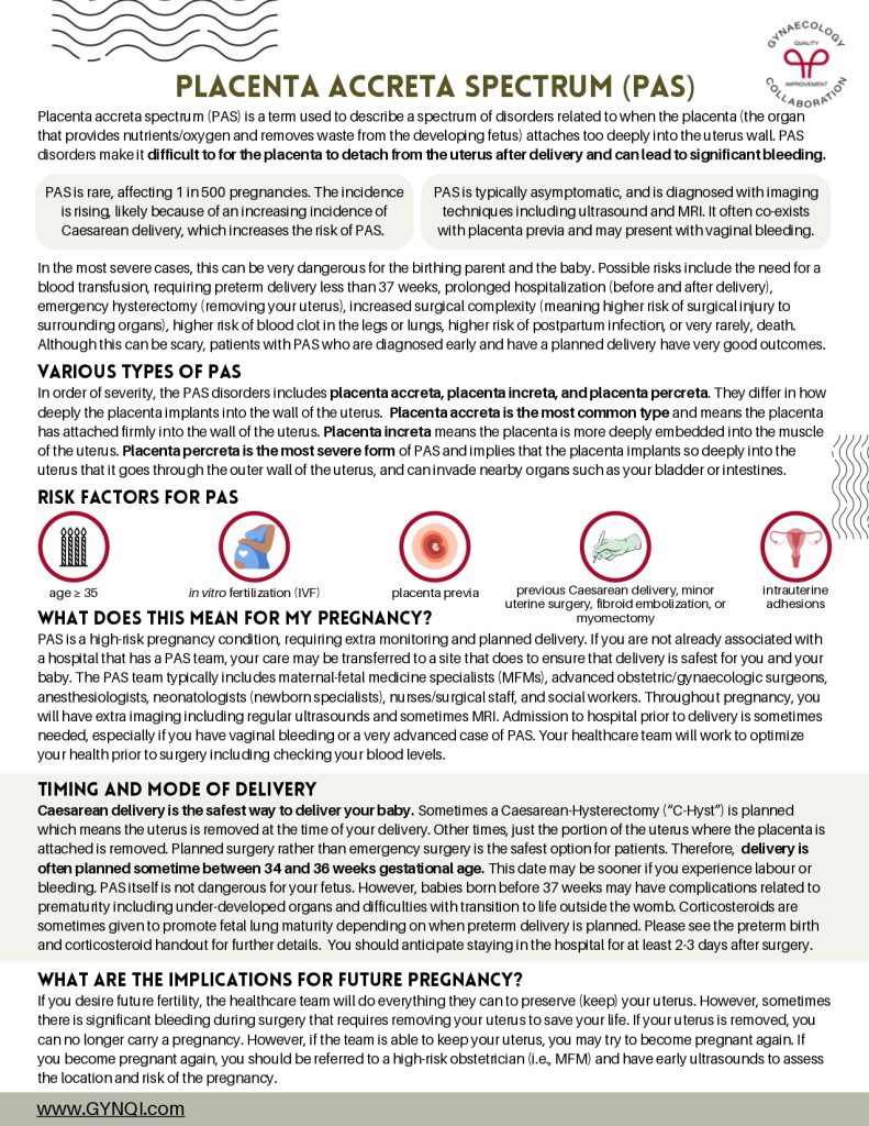
Risk Factors for PAS
- Age ≥35
- In vitro fertilization (IVF)
- Placenta previa
- Previous Caesarean delivery, minor uterine surgery, fibroid embolization, or myomectomy
- Intrauterine adhesions
What Does This Mean for My Pregnancy?
PAS is a high-risk pregnancy condition, requiring extra monitoring and planned delivery. If you are not already associated with a hospital that has a PAS team, your care may be transferred to a site that does to ensure that delivery is safest for you and your baby. The PAS team typically includes maternal-fetal medicine specialists (MFMs), advanced obstetric/gynaecologic surgeons, anesthesiologists, neonatologists (newborn specialists), nurses/surgical staff, and social workers. Throughout pregnancy, you will have extra imaging including regular ultrasounds and sometimes MRI. Admission to hospital prior to delivery is sometimes needed, especially if you have vaginal bleeding or a very advanced case of PAS. Your healthcare team will work to optimize your health prior to surgery including checking your blood levels.
Timing and Mode of Delivery
Caesarean delivery is the safest way to deliver your baby. Sometimes a Caesarean-Hysterectomy (“C-Hyst”) is planned which means the uterus is removed at the time of your delivery. Other times, just the portion of the uterus where the placenta is attached is removed. Planned surgery rather than emergency surgery is the safest option for patients. Therefore, delivery is often planned sometime between 34 and 36 weeks gestational age. This date may be sooner if you experience labour or bleeding. PAS itself is not dangerous for your fetus. However, babies born before 37 weeks may have complications related to prematurity including under-developed organs and difficulties with transition to life outside the womb. Corticosteroids are sometimes given to promote fetal lung maturity depending on when preterm delivery is planned. Please see the preterm birth and corticosteroid handout for further details. You should anticipate staying in the hospital for at least 2-3 days after surgery.
What Are the Implications for Future Pregnancy?
If you desire future fertility, the healthcare team will do everything they can to preserve (keep) your uterus. However, sometimes there is significant bleeding during surgery that requires removing your uterus to save your life. If your uterus is removed, you can no longer carry a pregnancy. However, if the team is able to keep your uterus, you may try to become pregnant again. If you become pregnant again, you should be referred to a high-risk obstetrician (i.e., MFM) and have early ultrasounds to assess the location and risk of the pregnancy.
Vasa Previa
In a normal pregnancy, the blood vessels that bring blood between your fetus and your placenta are protected by the umbilical cord or the placenta. Vasa previa is a very rare condition (1 in 1275-5,000 pregnancies) where unprotected blood vessels of the fetus run near or over the cervix. This is a problem because labour and/or breaking of the waters can cause the blood vessels to break, leading to devastating bleeding for your fetus. Vasa previa cannot be “cured” but diagnosis during your pregnancy allows us to manage and prevent serious consequences.
Diagnosis
Most patients with vasa previa have no symptoms. Vasa previa should be diagnosed on ultrasound. Rarely, it is diagnosed with vaginal bleeding or on physical exam in labour. Vasa previa may resolve and so the diagnosis is reconfirmed in the third trimester.
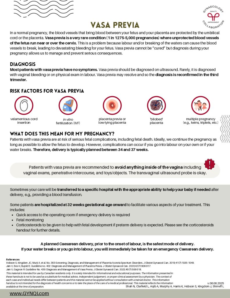
Risk Factors for Vasa Previa
- Velamentous cord insertion
- In vitro fertilization (IVF)
- Placenta previa or low-lying placenta
- ‘Bilobed’ placenta
- Multiple pregnancy (e.g., twins, triplets, etc.)
What Does This Mean For My Pregnancy?
Patients with vasa previa are at risk of serious fetal complications, including fetal death. Ideally, we continue the pregnancy as long as possible to allow the fetus to develop. However, complications can occur if you go into labour on your own or if your water breaks. Therefore, delivery is typically planned between 34 and 37 weeks.
Patients with vasa previa are recommended to avoid anything inside of the vagina including vaginal exams, penetrative intercourse, and toys/objects. The transvaginal ultrasound probe is okay.
Sometimes your care will be transferred to a specific hospital with the appropriate ability to help your baby if needed after delivery, e.g., providing a blood transfusion.
Some patients are hospitalized at 32 weeks gestational age onward to facilitate various aspects of your treatment. This includes:
- Quick access to the operating room if emergency delivery is required
- Fetal monitoring
- Corticosteroids to be given to help with fetal development if preterm delivery is expected. Please see the corticosteroids handout for further details.
A planned Caesarean delivery, prior to the onset of labour, is the safest mode of delivery. If your water breaks or you go into labour, you will immediately be taken for an emergency Caesarean delivery.